Yun Li1,
Fang Li1,2,
Hongyan Cai1,
Xuan Chen1,
Wei Sun1,
Wangyang Shen1,2 ![]()
For correspondence:- Wangyang Shen Email: whwangyangshen@126.com Tel:+862783924790
Received: 24 May 2016 Accepted: 14 September 2016 Published: 31 October 2016
Citation: Li Y, Li F, Cai H, Chen X, Sun W, Shen W. Structural characterization of inclusion complex of arbutin and hydroxypropyl-β-cyclodextrin. Trop J Pharm Res 2016; 15(10):2227-2233 doi: 10.4314/tjpr.v15i10.22
© 2016 The authors.
This is an Open Access article that uses a funding model which does not charge readers or their institutions for access and distributed under the terms of the Creative Commons Attribution License (http://creativecommons.org/licenses/by/4.0) and the Budapest Open Access Initiative (http://www.budapestopenaccessinitiative.org/read), which permit unrestricted use, distribution, and reproduction in any medium, provided the original work is properly credited..
Purpose: To improve the solubility and stability of arbutin and to expand its application by preparing its inclusion complex with hydroxypropyl-β-cyclodextrin (HP-β-CD).
Methods: An inclusion complex made of arbutin and hydroxypropyl-β-cyclodextrin (HP-β-CD) was prepared by freeze-drying method. Various analytical techniques, including ultraviolet-visible spectroscopy (UV), Fourier transform infrared spectroscopy (FT-IR), scanning electron microscopy (SEM), x-ray diffractometry (XRD) and thermo-gravimetric/differential scanning calorimetry (TG/DSC), were used to characterize the inclusion complex.
Results: UV spectra indicated that no new unsaturated bond was formed in the inclusion complex. Infrared analysis showed that the smaller peaks in the proximity of 1450 - 1600 cm-1 were characteristic of the aromatic nucleus, indicating that the phenyl ring of arbutin was involved in the formation of the inclusion complex. Scanning electron micrographs of the inclusion complex showed that the original morphology of both components disappeared, and some tiny aggregates of amorphous areas of irregular size were present, revealing that the arbutin was dispersed in HP-β-CD. The powder XRD pattern of the inclusion complex was more similar to that of amorphous HP-β-CD and did not exhibit the characteristic peaks of arbutin which suggest that arbutin in HP-β-CD matrix was molecularly dispersed, and existed in an amorphous state. The TG curve of the inclusion complex was a one-step process, partly proving the formation of the complex. Complex formation with HP-β-CD remarkably improved the physical and chemical stabilities of arbutin.
Conclusion: Inclusion complex of arbutin with HP-β-CD improves the heat stability of arbutin remarkably. This has a potential for expanding the application of arbutin to pharmaceuticals and food.
Introduction
Arbutin (4-hydroxyphenyl-α-D-glucopyranoside) is an important phenolic compound found in many plants and fruits [1]. It has potent antifungal, antiviral and anticancer activities [2-4]. Arbutin was initially identified as an antibiotic substance in fire blight resistance [5], and later as a specific marker for evaluation of pear product authenticity [6]. It has also attracted attention for its antitussive and antibacterial effects. Studies have shown that arbutin has antioxidant properties [7-9] and is rich in phenolic compounds [9,10]. Arbutin inhibits biosynthesis of the human pigment melanin, which is the basis of its application in skin-lightening cosmetics [11,12]. Arbutin is also used to treat infections of the urogenital tract [13].
However, the poor aqueous solubility and poor stability of arbutin in water has restricted its application. Water solubility of natural compounds is generally improved by glycosylation using enzymatic or chemical methods. Glycosylation is an important process used to structurally modify biologically active phenolic compounds, so as to render them more soluble in aqueous systems and increase their stability to light and oxidation. Attachment of a sugar moiety to a molecule increases its hydrophilicity and improves its physicochemical and pharmacokinetic properties [14]. However, a water-soluble polymer containing glucose residues may cause biological responses when it is metabolized. The glycosyl structures are appropriately modified [15]. Animal studies have shown that arbutin metabolites have hepatotoxic, nephrotoxic, mutagenic and carcinogenic potential [16,17]. Thus, glycosylation may create a group of new compounds with unknown safety, making it necessary to ascertain their safety before their application in food or medicines [18].
Cyclodextrins (CDs) are non-toxic macrocyclic oligosaccharides consisting of (α-1, 4)-linked α-L-glucopyranose units. With hydrophilic outer surfaces and hollow hydrophobic interiors, CDs are very attractive ingredients for making artificial enzymes and other biomimetic materials. They are readily available. They can bind hydrophobic substrates into their cavities in aqueous solutions, and they have two rows of hydroxyl groups that can either react with substrates themselves or be widely used to attach other catalytic functional groups. Consequently CDs are widely used in the food industry as food additives, for stabilization of flavors, for elimination of undesired tastes or other undesired compounds such as cholesterol and for prevention of microbiological contaminations and browning reactions [19,20]. The most common CDs used as formulation vehicles are α-, β- and γ-CDs containing six, seven and eight glucopyranose units, respectively.
Hydroxylpropyl-β-cyclodextrin (HP-β-CD), a hydroxyalkyl derivative, is an alternative to α-, β- and γ-CDs. It has improved water solubility and may be more toxicologically benign [21,22]. It has been confirmed that HP-β-CD is well tolerated in humans, the main adverse event being diarrhoea [23].
To the best of our knowledge, there are no reports on preparation of an inclusion complex of arbutin and HP-β-CD. In this study, we prepared an inclusion complex of arbutin with HP-β-CD for the first time, studied its physicochemical properties and characterized it by ultraviolet-visible spectroscopy (UV), Fourier transform infrared spectroscopy (FT-IR), scanning electron microscopy (SEM), X-ray diffractometry (XRD) and thermogravimetric/differential scanning calorimetry (TG/DSC).
Methods
Materials and chemicals
Arbutin (> 99 %) was obtained from Shanxi Huike Botanical Development Co., Ltd., (Xi’an, China). Hydroxypropyl-β-cyclodextrin (MW 1460) was obtained from Sigma Chemical Co. (USA). All other chemicals and solvents were of analytical grade.
Preparation of the inclusion complex of arbutin and HP-β-CD
The complex was prepared by mixing arbutin and HP-β-CD, 1:1 molar ratio, using freeze-drying method. Arbutin (0.272 g) and HP-β-CD (1.460 g) were dispersed in 50 mL of distilled water; the mixture was kept stirred for 72 h at 40 °C, and then filtered. The filtrate was freeze-dried, and to yield the powdery inclusion complex, which was collected for the physicochemical analysis.
Preparation of physical mixture of arbutin and HP-β-CD
Arbutin and HP-β-CD were separately pulverized in ceramic mortars. Thereafter, arbutin and HP-β-CD were mixed at a molar ratio of 1:1 in a spatula to obtain l a homogeneous mixture.
UV-visible spectroscopy (UV)
UV spectra were recorded for arbutin, HP-β-CD, their physical mixture and inclusion complex using a TU-1810PC spectrophotometer (Beijing Purkinje General Instrument Co., Ltd., Beijing, China). Each sample was dissolved in distilled water at room temperature. The aqueous solutions were scanned in the wavelength range of 200 to 400 nm to obtain the UV spectra.
Fourier transform infrared spectroscopy (FT-IR)
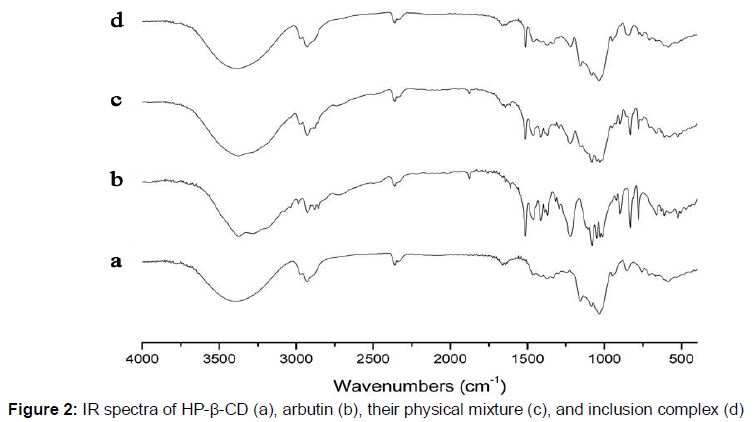
IR spectra of arbutin, HP-β-CD, their physical mixture and inclusion complex were obtained between 4000 and 400 cm-1 (Mid infrared region) on a TENSOR 27 infrared spectrophotometer (Bruker, Germany) with 256 scans at a resolution of 4 cm-1. Each sample was ground with spectroscopic grade potassium bromide (KBr) powder and then pressed into 1 mm pellets (2 mg of sample per 200 mg dry KBr). A blank KBr disk was used as background/control.
Scanning electron microscopy (SEM)
Surface morphologies of arbutin, HP-β-CD, their physical mixture and inclusion complex were investigated using a Quanta 200 environmental scanning electron microscope (FEI, USA). Prior to examination, the samples were prepared by mounting about 0.5 mg of powder onto a 5 mm × 5 mm silicon wafer affixed via graphite tape to an aluminum stub. Then the samples were examined at the accelerating potential of 15 kV under low vacuum.
X-ray diffractometry (XRD)
Monochromatic Cu Ka radiation (wavelength = 1.54056 A˚) was produced by a D8 X-ray diffractometer (Bruker, Germany). Each sample powder was packed tightly in a rectangular aluminum cell. The samples were exposed to the x-ray beam from an x-ray generator. The scanning regions of the diffraction angle, 2θ, were 5 – 80˚. Duplicate measurements were made at ambient temperature. Radiation was detected with a proportional detector.
Thermogravimetric/differential scanning calorimetry (TG/DSC)
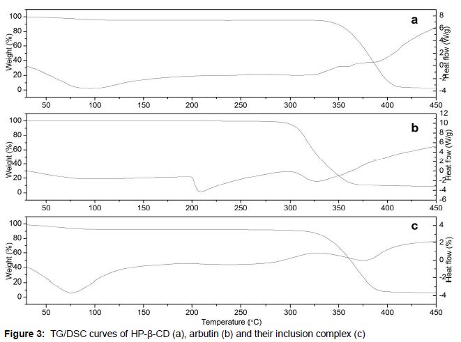
Q600 TG/DSC system (TA, USA) was adjusted to operate in dynamic atmosphere of nitrogen (99.999 %) at 100 mL/min and heating rate of 20 °C/min from 20 to 500 °C, with a sample mass of about 5 mg.
Results
The absorption spectra of arbutin, HP-β-CD, their physical mixture and inclusion complex are presented in . Due to the absence of double bond, there is no absorption peak for HP-β-CD. However, two remarkable absorption peaks were found at wavelengths of 221 nm and 283 nm, which were confirmed as the characteristic absorption peaks of arbutin.
The infrared spectra of arbutin, HP-β-CD, their physical mixture and inclusion complex are shown in . For HP-β-CD in this IR spectrum, several prominent absorption bands were found at 3403 cm-1 (for O-H stretching vibrations), 2930 cm-1 (for C-H stretching vibrations) and 1157, 1084 and 1033 cm-1 (for C-H, C-O stretching vibration). The FT-IR spectrum of arbutin consisted of the prominent absorption bands of the hydroxyl group (3373cm-1) and the aromatic nucleus (1512, 1461, 1411 cm-1). It is interesting that an additive effect of arbutin and HP-β-CD was displayed in both the IR spectra of the physical mixture and the inclusion complex. Although all the characteristic absorption peaks of arbutin and HP-β-CD could still be found, the spectrum of the complex was mainly affected by the HP-β-CD, in disappearance of some characteristic absorption peaks of arbutin in the wavelength bands between 400 and 1500 cm-1. The smaller peaks in the proximity of 1450 - 1600 cm-1 are characteristic of the aromatic nucleus, indicating that the phenyl ring of arbutin was involved in the formation of the inclusion complex.
The surface morphology of the powders derived from arbutin, HP-β-CD, their physical mixture and complex was assessed by SEM. As illustrated in Plate 1, HP-β-CD was observed as amorphous spheres (Plate 1a), whereas the arbutin existed in the form of needle-like crystals (Plate 1b). Both characteristics, amorphous spheres and needle-like crystal, were found in the physical mixture of HP-β-CD and arbutin (Plate 1c). As for the inclusion complex, the original morphology of both components disappeared and some tiny aggregates of amorphous pieces with irregular size were present (Plate 1d).
Powder x-ray diffraction patterns of arbutin, HP-β-CD, their physical mixture and inclusion complex are shown in . One broad peak was observed for the HP-β-CD (Plate 2a), while some very sharp crystalline peaks were shown in the powder diffraction pattern of arbutin (b). As shown in Plate 2c, a superimposition of the patterns of the two compounds, arbutin and HP-b-CD, was very distinct. However, the powder XRD pattern of the inclusion complex was more similar to that of the amorphous HP-β-CD and did not exhibit the characteristic peaks of arbutin (d).
TG and DSC curves of arbutin, HP-β-CD and inclusion complex are shown in Plate 3. It was found that with increase in temperature, HP-b-CD began to decompose at about 320 °C and then ended at about 420 °C. The decomposing peak value (about 360 °C) was obtained from the DSC curve (a). The fusion and degradation of HP-b-CD were completed in one step. Different from this, the DSC curve of arbutin had an obvious endothermic peak where the mass of arbutin did not decrease (b), suggesting that the arbutin melted at about 200 °C. Therefore, the degradation of arbutin began at about 200 °C, and ended at about 370 °C (Plate 3b). The TG curve of the inclusion complex was also a one step process (Plate 3c), partly proving the formation of the complex. The starting temperatures for mass loss were about 320°C, ended at about 420 °C (c).
Discussion
UV spectroscopy is an important and efficient tool for studying the formation of complex of arbutin and HP-β-CD. The curves of physical mixture and inclusion complex of arbutin and HP-β-CD did not coincide, which indicates that the inclusion complex of arbutin and HP-β-CD was not a simple physical mixture. The characteristic absorption peaks of their inclusion complex were not changed, indicating no new unsaturated bond was formed.
Infrared analysis showed that the infrared spectrum of the physical mixture of arbutin and HP-β-CD was the superimposition of arbutin and HP-β-CD. However, the infrared spectrum of the inclusion complex of arbutin and HP-β-CD were similar to that of arbutin. There were no obvious new absorption peaks in the inclusion complex, indicating that there was no new chemical bond formation during the inclusion complex formation process.
Scanning electron microscope captures the surface morphology of a sample by the interaction between electrons and the sample material. The two characteristics, amorphous spheres and needle-like crystal, were found in the physical mixture of HP-β-CD and arbutin. As for the inclusion complex, the original morphology of both components disappeared, and some tiny aggregates of amorphous pieces with irregular size were present, revealing that the arbutin did not exist in the crystal state but was dispersed in HP-β-CD.
X-ray diffractometry is a very useful method for the detection of CDs complexes in powder or microcrystalline states. The diffraction pattern of the complex is supposed to be clearly distinct from that formed from mere superposition of the components if a true inclusion complex is formed.
The diffraction pattern of arbutin which exhibited many spikes indicated that it was crystalline, while HP-β-CD showed no spikes, indicating that it is in amorphous state. The physical mixture of arbutin and HP-β-CD could decrease the characteristic crystallization peak of arbutin to some extent. The inclusion complex showed no crystallization peak of arbutin due to the effect of HP-β-CD. X-ray diffraction analysis suggested that arbutin in the HP-β-CD matrix was molecularly dispersed, and existed in an amorphous state.
Thermogravimetric (TG) and differential scanning calorimetry (DSC) are important methods for research on thermal properties of cyclodextrin and its inclusion complex. Thermogravimetric -differential scanning calorimetry combined with (DSC-TG) is the latest development in thermal analysis technology, which can simultaneously test weight changes and heat absorption/heat release of samples with increase in temperature. The DSC curve of arbtin shows one endothermic peak at 320 °C. However, this characteristic endothermal peak disappeared in the DSC curve of the inclusion complex. This shows that arbutin was completely dispersed in HP-β-CD and that there was some kind of interaction between them, such as hydrogen bonds or van der Waals force [24]. The decomposition temperature of the complex was about 300 °C, indicating that the heat stability of arbutin could be remarkably improved by forming an inclusion complex with HP-β-CD.
Conclusion
The findings of this study show that the arbutin in the inclusion complex is completely dispersed in HP-β-CD matrix. It is worth noting that arbutin in the HP-β-CD matrix is molecularly dispersed without forming a new unsaturated bond. A combination of hydrogen bonds or van der Waals forces may be responsible for the formation of the inclusion complex. Formation of inclusion complex with HP-β-CD can improve the heat stability of arbutin remarkably, and enhance its application in the field of pharmaceuticals and food.
Declarations
Acknowledgement
References
Archives


News Updates